

Bone: The Clavicle
Type of Bone: It’s a Long bone, with concave and convex curvature.
Structure: The Bone consist of the Shaft, Medial End, and Lateral End
The Anatomy of the Clavicle Bone
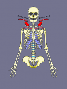
We are looking at the right clavicle bone, from an anatomical position.
The clavicle bone is also known as the collarbone. It’s in the family of long bones, it’s the only long bone that lays horizontal in the human body, every other long bone is vertical.
The Clavicle Bone can be divided into 3 sections. On the lateral end of the bone we have the Acromial End. And on the medial end, we have the Sternal end. The Sternal end connects to the Manubrium of the Sternum and the Acromial end connects to the acromion of the scapula.
In the middle we have the shaft, the body of the Clavicle and that shaft is divided into 2 parts . The lateral ⅓ rd’s and medial ⅔ rd’s. The lateral end is flat and the medial end is round.
Both the Lateral end and medial end have 2 surfaces and 2 borders, the lateral 1/3rd surfaces are referred to as the upper surface and lower surface. Whereas the medial end is referred to as the Superior and Inferior surface. Both the lateral and medial sides of the shaft have an Anterior and posterior border.
The superior portion of the bone is mostly smooth as a baby’s bottom and the inferior portion is as rough. The anterior border of The medial end of the clavicle is Covex and The lateral end is concave.
Now let’s move onto the Bone Anatomy, we will start on the lateral end “The Acromial end” and work our way medialy, On the Acromial end we have the Acromial Facet. This facet articulates with the acromion of the Scapula.This makes up the acromioclavicular joint.
First bony landmark is the Trapezoid line, it provides an attachment site for the Trapezoid ligament. Next up is the Conoid tubercle, it’s located here and it provides an attachment site for the conoid ligament. Those two ligaments we just mentioned, are part coracoclavicular ligament.
Next up is the Subclavian groove, It’s an actual groove in the bone, and it is where the Subclavious muscle inserts. On the lateral portion of the Subclavian groove is where the Nutrient Foramen is, it provides all the nutrients to make a strong healthy bone, through a (nutrient artery).
In fact the Clavicle is the first bone to start growing when you’re in mommy’s belly and one of the last bones to stop growing, 21 to 25 years old.
Next up we have the impression for the costoclavicular ligament, and as the name says this is where that ligament attaches. Now don’t confuse this with the Coracoclavicular ligament, this is the costoclavicular ligament Costo-meaning ribs.
Now we have arrived at the medial end, the sternal end. Located here is the sternal facet which articulates with the Manubrium of the sternum to make the sternoclavicular joint.
Bone Anatomy
Muscle Attachments
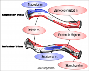
Origins:
- Sternocleidomastoid Muscle
- Deltoid Muscle
- Pectoralis Major muscle
- Sternohyoid Muscle
Insertions:
- Trapezius Muscle
- Subclavius
Fun Facts
- Though it is a Long Bone, it does not have bone marrow like other long bones
- The Clavicle is the most commonly fractured bone in the human body.
QUIZ & ANSWERS
The Quiz and and Answer Key are below
Download the .pdf for free below